10+ diagram of replication fork
DNA replicates in a semi-conservative manner in which each individual strand is copied to form a new molecule of DNA. To do this a particular protein called single-stranded DNA binding proteins SSBs covers and keeps the isolated strands of DNA close to the replication fork.
Browse Questions For Biology
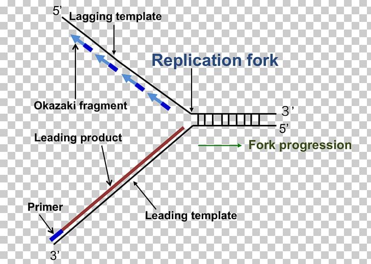
It forms at the repication bubble with the help of the enzyme DNA helicase.

. The two strands can be labelled with isotopes using substrates that. National Library of Medicine. The proliferation of all organisms depends on the coordination of enzymatic events within large multiprotein replisomes that duplicate chromosomes.
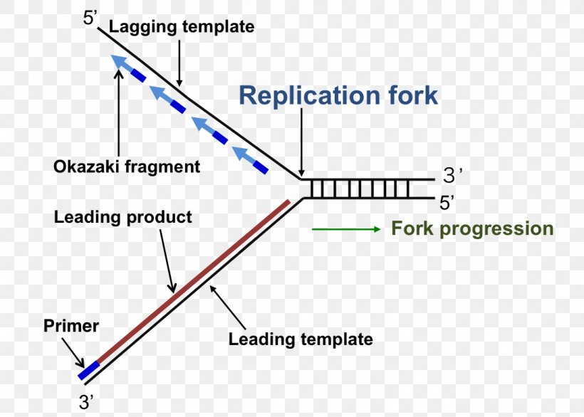
When a cell divides during mitosis the first thing it has to do is ensure that the new cells will have identical DNA to itself. Label the components first blue and then the. In the following diagram of a replication fork which DNA strand a or b is a.
What is the purpose of a replication fork. It has two branching prongs each of which is made up of. DNA replicates in a semi-conservative manner in which each individual strand is copied to form a new molecule of DNA.
The template for the synthesis of the lagging strand. 8600 Rockville Pike Bethesda MD 20894 USA. National Institutes of Health.
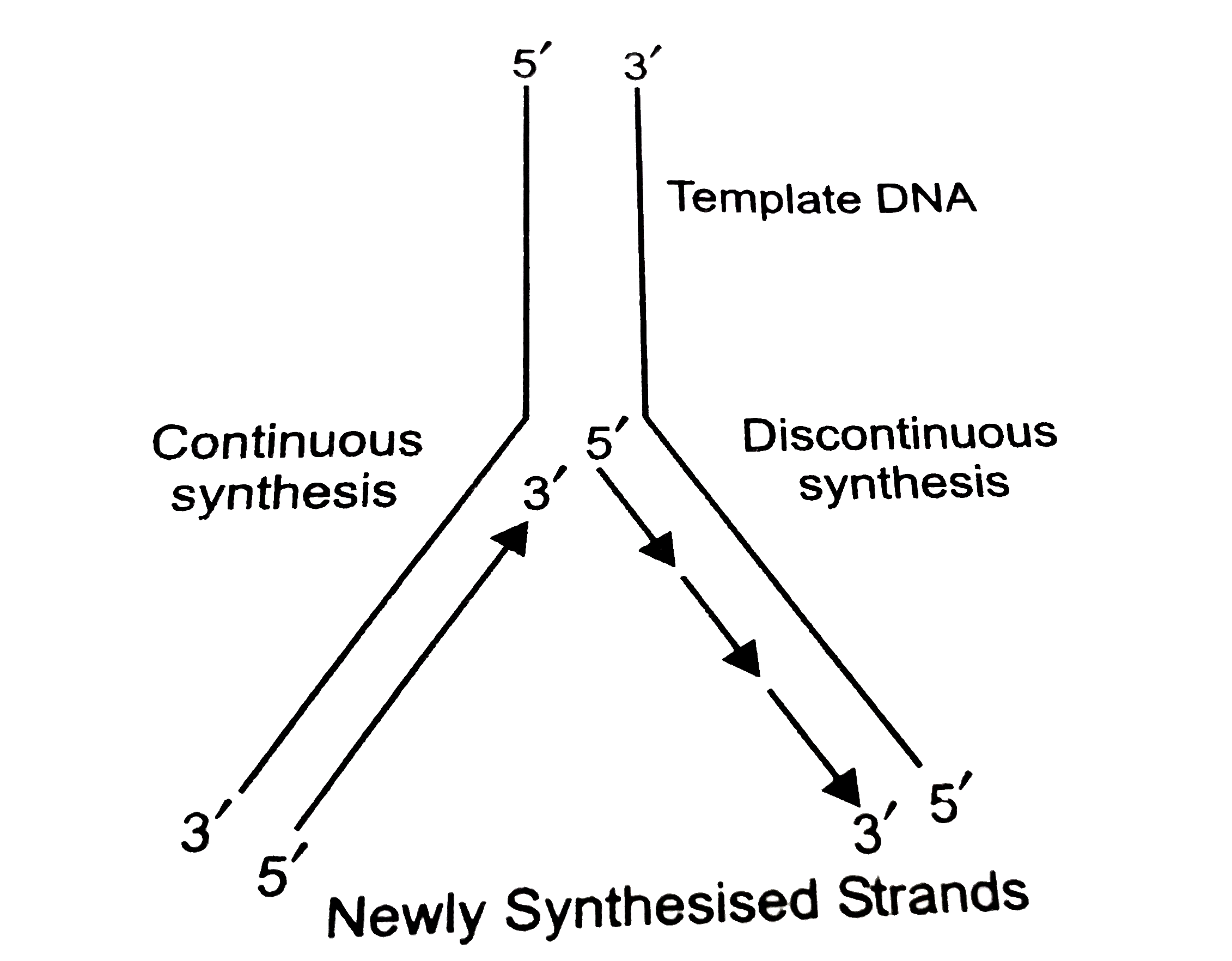
This leads to two strands called leading and lagging strands. The replication fork is a region where a cells DNA double helix has been unwound and separated to create an area where DNA polymerases and the other enzymes involved can use each strand. 1 mark Direction of fork movement 5 b.
The leading strand is directed from 3 to 5 The lagging strand is orientated from 5 to 3 The two sides are reproduced using two distinct methods to account. Replicating fork is the structure of the DNA double helix after the unzipping by ligase enzyme. The replication fork is two-way.
The replication fork is a Y-shaped structure. Start studying Replication Fork Diagram. Replication begins at an origin of replication where two strands are separated opening a replication bubble.
The two strands can be labelled with isotopes using substrates that. Learn vocabulary terms and more with flashcards games and other study tools. Complete diagram of a replication fork in bacterial DNA Drag the appropriate labels to their respective targets.
The replication fork is a structure that is opened by DNA helicase within the long helical DNA during DNA replication. A eukaryotic chromosome may have hundreds or thousands of origins of. The replication of DNA occurs during the S.
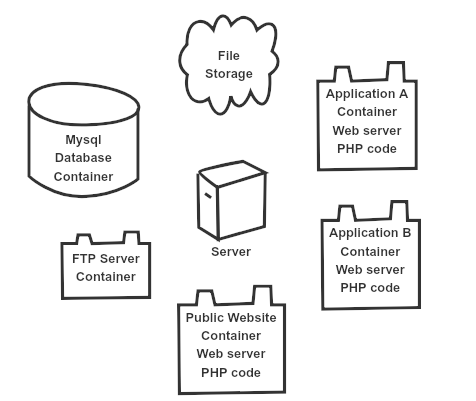
Clustered Web Applications Mysql And File Replication Roojsolutions
Draw A Labelled Schematic Sketch Of Replication Fork Of Dna Explain The Role Of The Enzymes Involved In Dna Replication
Dna Replication Replication Fork Enzyme Triangle Png Clipart Angle Diagram Dna Dna Replication Enzyme Free Png

Approaches To Heterogeneity In Native Mass Spectrometry Chemical Reviews

What Is The Ligase S Function In Dna Replication Quora
Draw A Labelled Diagram Of Replicating Fork
Web3 Is Just Expensive P2p R Programming

Biological Mechanisms And Clinical Significance Of Bap1 Mutations In Human Cancer Abstract Europe Pmc
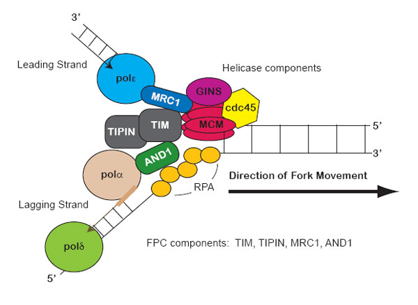
Replication Fork Components Learn Science At Scitable
Frontiers Genetic Regulation Of Vertebrate Forebrain Development By Homeobox Genes

Commit Latency Is Fluctuating And Too High Issue 8551 Camunda Zeebe Github

Senescence In Yeast Is Associated With Chromosome Xii Cleavage Rather Than Ribosomal Dna Circle Accumulation Biorxiv

Draw A Labelled Schematic Sketch Of Replication Fork Of Dna Explain The Role Of The Enzymes Involved In Dna Replication

Dna Replication Wikiwand
Phosphorylation Of The Mbf Repressor Yox1p By The Dna Replication Checkpoint Keeps The G1 S Cell Cycle Transcriptional Program Active Plos One

Draw A Labelled Diagram Of Replicating Fork
Dna Replication Replication Fork Enzyme Triangle Png 1101x788px Dna Replication Diagram Dna Enzyme Parallel Download Free